Skip to the 3D experience
JINTAI3D

Made possible by open-source anatomy.

Explore anatomy.All for free.

Anatomy data shared by researchers, brought closer to everyday learning. The JINTAI 3D series uses open-source anatomy models. JINTAI 3D Model, Challenge, Book and NeuroMap are all free to use. Rotate, separate and discover how the body connects.

Open JINTAI 3D Model for freeNeuroMap is now available

No sign-up, payment or installation. Just your browser.
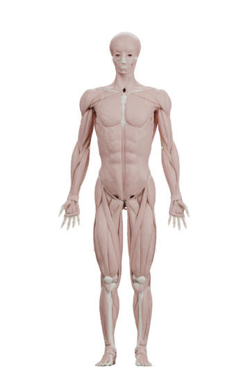
Z-Anatomy model showing the muscles and bones of the human body
Drag or use the arrow keys to rotate the anatomical model. On a phone, swipe horizontally to rotate.Loading the 3D specimen
An actual Z-Anatomy model
Drag to rotate
Try the interactive model

Beyond knowing the names.See where things are.Discover how they connect.

The overlap of muscles. Their position around a bone. Things a single illustration can make hard to see. Change your perspective and explore at your own pace. The JINTAI 3D series is made for those small discoveries.

A 3D model of the human body
Drag to rotate

One human body.So much to discover.

Switch between the views below and discover anatomy in three dimensions.

Look beyond the overlap.

Separate bones and muscles into individual structures. Open up their overlapping layers and connect the whole body with its parts.

The exploded view moves structures from their anatomical positions to make them easier to see.

Open JINTAI 3D Model for free

Actual BodyParts3D data

Beneath the muscles.A world within.

Organs sheltered by bone. Structures woven together. Explore the inside of the body from your own perspective.

Open the whole-body viewer
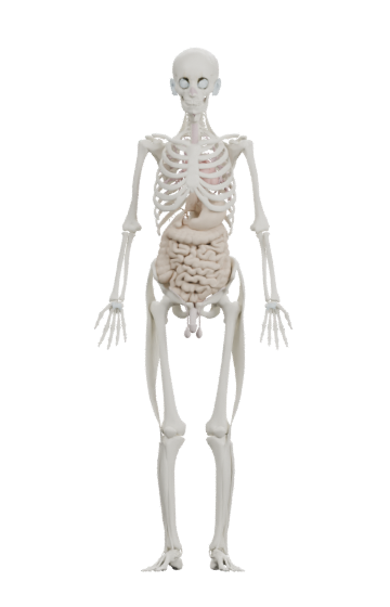
Whole-body model with internal organs

Get closer to the organs that interest you.

A view beneath the skeleton.Drag to rotate
0%
Scroll to look insideCC BY 4.0

The exploded view moves structures from their anatomical positions.

The desire to understandthe body has a long history.

Drawn on paper. Modeled in wax. Shared as data. The way we teach anatomy keeps evolving.

  1. 1543

    The body, on paper.

    Vesalius publishes the Fabrica, using detailed anatomical illustrations to communicate the structures of the human body.

    US National Library of Medicine
  2. 18th c.

    Knowledge takes shape.

    Anatomical wax modeling flourishes in Florence, giving teaching materials a physical form and a third dimension.

    University of Florence
  3. 2007

    Anatomy becomes open data.

    BodyParts3D is released in Japan, providing a shared foundation for the shapes and positions of human body structures.

    Database Center for Life Science
  4. Today

    The next chapter is in your hands.

    Open models, made accessible in a browser. The JINTAI 3D series brings anatomical knowledge closer to the people learning it.

    Explore our model sources
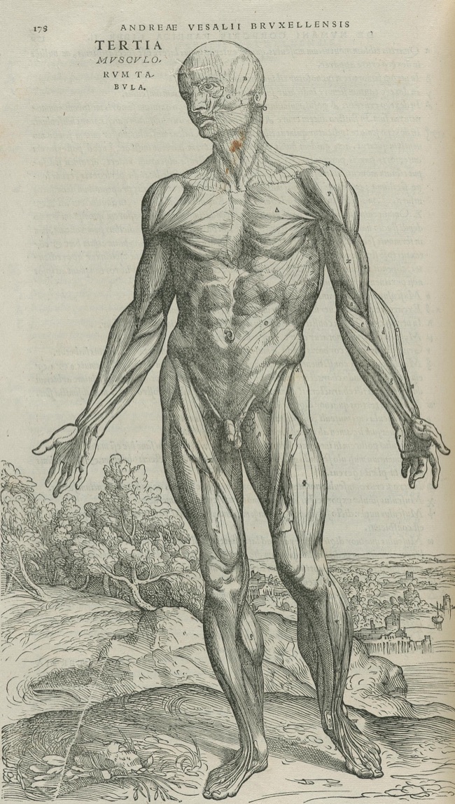
A woodcut of the muscles from Vesalius’s 1543 Fabrica.
Andreas Vesalius, 1543.
Courtesy of the National Library of Medicine.
Image credits

For people whose knowledgemakes a difference to others.

Anatomy for your everyday.

A shared view of the body.

Some relationships are difficult to describe in words alone. Rotate the model and reveal structures to create a shared visual reference when talking about the body.

Open JINTAI 3D Model for free

An open doorto understanding anatomy.

Specialist learning should be easier to access and afford. We build on anatomy models that the world already shares.

The JINTAI 3D series uses open data from BodyParts3D and Z-Anatomy. By crediting the people behind these resources and preserving their licenses, we connect their work with a browser-based learning experience.

No room for a physical model? No specialist software?

All you need to get started is a browser. Open the current learning tools and see how they fit the way you study.

Built on shared knowledge.

Follow your curiosity.Find your way in.

Explore with your hands. Put your knowledge to the test. Take time with each structure. Four ways into anatomy.

Explore & understand

JINTAI 3D Model

Find it. Turn it.Make the connection.

Muscles, bones, and organs. Rotate the body, isolate a structure, and open up the layers. Keep what you want to see and explore relationships a flat diagram can make hard to follow.

  • Search by name
  • Isolate a structure
  • Separate the layers
Open JINTAI 3D ModelStart with the organs
An actual view of JINTAI 3D Model3D credits

Try & discover

JINTAI 3D Challenge

You’ve seen it.Can you name it?

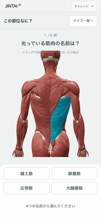
Touch a muscle. Recognize a shape. Find a structure beneath the layers. Turn observation into a challenge and discover what you know, five questions at a time.

  • Touch
  • Name
  • Shape
  • System
  • Left or right
  • Peel & find

Six ways to play. Five questions per challenge.

Try JINTAI 3D Challenge
Try one question here

Try one question. What is this bone?

Preparing the specimen…

Drag to rotate · Or use the arrow keys

Z-Anatomy · Model source · CC BY-SA 4.0

Actual mobile screen · Try it in the app

Read & go deeper

JINTAI 3D Book

Anatomy comesoff the page.

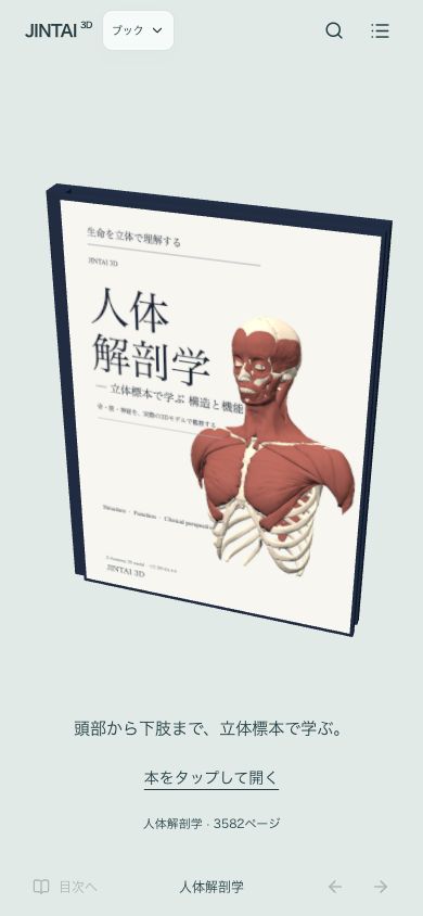
A 3,582-page atlas, from head to lower limb. Search for a structure, turn its specimen around, and see how it sits within the body. Make time to get to know each part.

  • Japanese & English name search
  • Rotate & enlarge specimens
  • See surrounding structures
Open JINTAI 3D Book
Actual screens from JINTAI 3D Book3D credits
3,582pages to explore

From head to lower limb. From one structure to the whole body.

Trace sensory regions

JINTAI 3D NeuroMap

Explore sensation.
In three dimensions.

Rotate and zoom the body to explore dermatomes, including the back, arms and legs that a single flat view cannot show.

  • Rotate and zoom
  • Learning and reference views
  • Sensory test points
Open NeuroMap

An educational preview, pending medical review. Not for diagnosis.
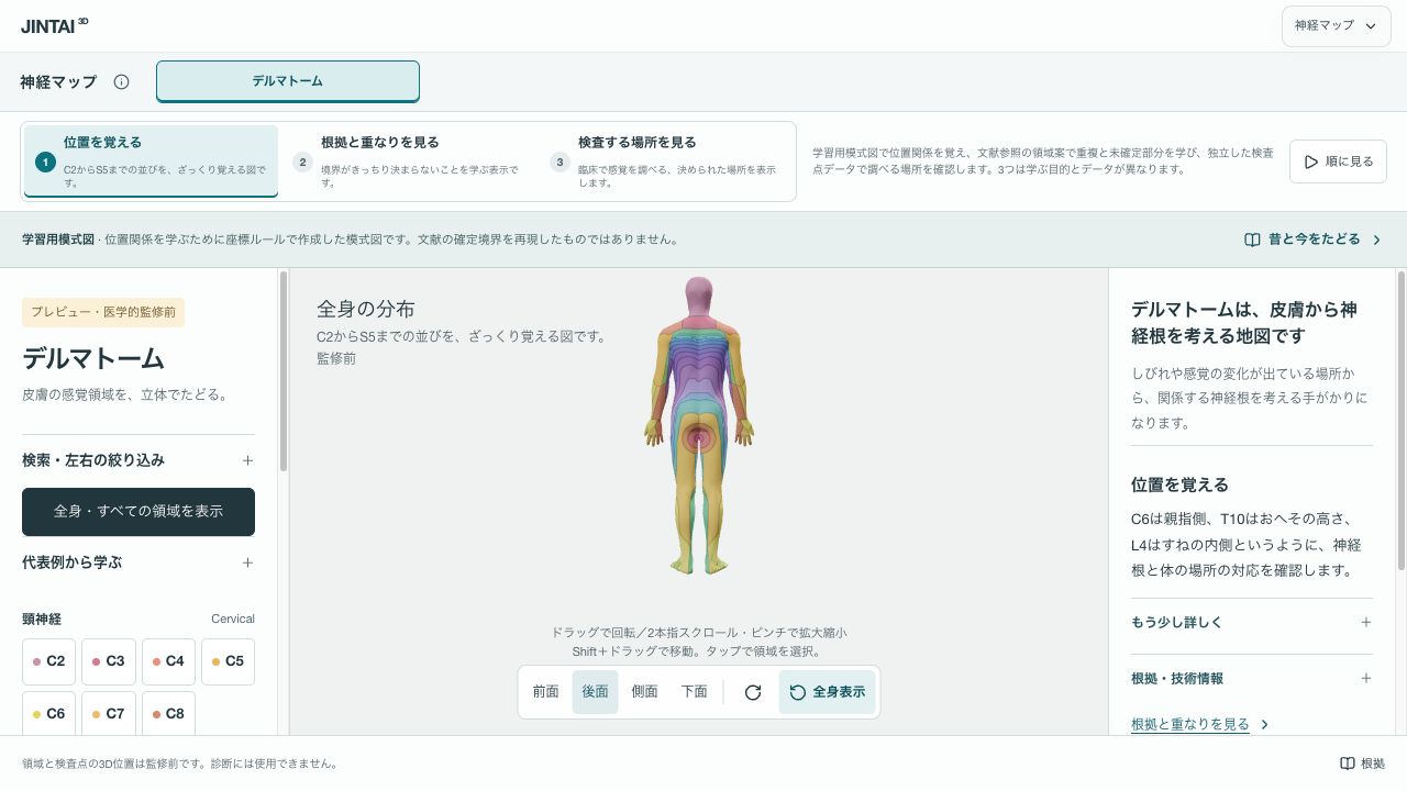
The NeuroMap interface
Live NeuroMap · Rotate to explore

Where will youstart today?

All four experiences open without registration.

The JINTAI 3D Book and JINTAI 3D Challenge learning interfaces are primarily in Japanese.

Before you begin.

How much does it cost?

JINTAI 3D Model, JINTAI 3D Book, JINTAI 3D Challenge, and NeuroMap can currently be opened without registration or payment. Future paid plans and pricing have not been set. Our aim is to make anatomical learning easier to access.

Can I use it on my phone?

Use a browser that supports WebGL. The interface adapts to phones, tablets, and computers. Loading 3D models requires an internet connection; performance depends on your device and connection.

Which languages are available?

This page and the JINTAI 3D Model core interface are available in Japanese and English. Some explanations and learning features are primarily in Japanese. Some model labels remain in English.

Can I use it for diagnosis or treatment?

This is an educational anatomy reference, not a tool for diagnostic, treatment, or surgical decisions. The models do not cover every structure or individual variation, and anatomical accuracy in this application has not been fully reviewed by specialists. Some explanatory content was created with AI assistance.

A world of anatomy.In your hands.

Let today’s small discovery become tomorrow’s understanding.

Open JINTAI 3D Model for free

Updates

Follow new features, model additions and changes awaiting release.
  1. Published

    JINTAI 3D NeuroMap is now available

    • Explore dermatomes in 3D. Rotate and zoom the body, and switch between learning diagrams, reference views and sensory test points.
    • Updated the schematic regions of the shoulders, trunk, lower legs and buttocks.
    • An educational preview, pending medical review. Not for diagnosis.
  2. Published

    Added 19 shoulder ligament and thoracic node shapes

    • Added 14 bilateral shoulder ligaments and five thoracic node groups, bringing the unified atlas to 2,551 shapes.
    • Deferred one candidate with a source name/location conflict. Ligament attachments are not certified; lymphatic vessels and drainage paths are absent.
  3. Published

    Added 30 spinal cord segments

    • Registered 30 HRA male spinal cord surface segments to the existing spinal canal, bringing the unified atlas to 2,532 shapes.
    • Preserved source segmentation edges. Roots, cauda equina, coccygeal segments and gray/white subdivisions are absent; continuity with the medulla or peripheral nerves is not certified.
View all earlier updates
  1. Published

    Added 73 nerve, joint, lymph node and gland structures to the unified model

    • Added both sides of cranial nerves IX–XII and the sympathetic trunks, five thyroid/parathyroid structures, 22 joint structures and 36 lymph node groups. The unified model now contains 2,502 shapes.
    • Documented different left/right vagus coverage and source gaps. Lymphatic vessels are not included; missing source shapes were not generated.
    • Available in the unified model and recorded in the landing page updates. Physical iPhone/Safari testing remains pending.
  2. Published

    Five-question muscle challenge

    • Added a fixed five-question Z-Anatomy surface-muscle challenge.
    • Start after a countdown and total answer time only. Correct answers highlight the muscle; wrong answers do not hide parts.
    • Show per-question and total times, share/copy results and link to the same course. Interrupted runs are labeled practice.
  3. Published

    OpenSim JAM knee playback

    • Added right passive knee flexion using Lenhart 2015 bones/cartilage and published simulation results, approximately 0–90 degrees.
    • Replay tibial and patellar rotations and translations without the previous circle or separation correction.
    • Added time scrubbing, end stop, restart, close-up and source/validation details.
    • Other joints remain Z-Anatomy prototypes. Motion mode is accessible from either atlas.
  4. Published

    Simplified joint motion controls

    • Removed fixed-axis comparison, separation correction and axis controls; retained roll/glide as the default for elbows and knee flexion.
    • Grouped playback, reset, zoom and explanation controls; moved documentation into an on-demand sheet.
    • Start with a joint close-up and list only implemented motions. Other joints remain labeled as basic prototypes.
  5. Published

    Animated return from focus

    • Added a 0.65-second zoom-out when leaving focus, with interaction cancellation and reduced-motion support.
  6. Published

    Region navigation and selected names

    • Added smooth camera navigation to shoulders, waist, knees and feet in both models.
    • Show selected names above the action buttons without opening explanations.
  7. Published

    Clarified Japanese drag instructions

    • Standardized left/right click-and-drag wording in the desktop guide.
  8. Published

    Renamed the site to JINTAI 3D and moved its URL

    • Updated the opening, menu and browser title to JINTAI 3D.
    • Moved to jintai-3d.pages.dev with redirects from human-atlas.pages.dev.
  9. Published

    Basic hip, shoulder and neck motions

    • Added hip/shoulder flexion-extension and neck flexion-extension/lateral bending; verified ankle dorsiflexion/plantarflexion.
    • Use eight provisional cervical segments. Shoulder flexion is limited to a 120-degree test range while retaining the separate 180-degree reference.
    • Group available motions first and improve joint close-up viewpoints.
  10. Published

    Knee rolling/sliding test and joint close-up

    • Added an ideal-circle model for left/right knee flexion with a fixed femur, representing an unloaded configuration.
    • Added joint close-up and whole-limb views, with separate elbow/knee parameters.
    • Not fitted to real joint surfaces; compartment differences, axial rotation, menisci and patellar coupling remain unmodeled.
  11. Published

    Experimental elbow rolling and sliding geometry

    • Compare fixed-axis rotation with an ideal tangent-circle model for both elbows.
    • Optional virtual contact circles and mathematical instantaneous axes, with adjustable uncalibrated parameters.
    • Not fitted to bone surfaces or measured trajectories. Legacy separation is not applied.
  12. Published

    Disabled visual position correction by default

    • Keep position correction off by default and retain it for comparison. Rotation-axis calibration remains incomplete.
  13. Published

    Optional provisional rotation axes

    • Toggle faint reference and translated axes, distinguished by color and line style.
    • Toggle visual position correction for comparison. These axes are not measured instantaneous joint axes.
  14. Published

    Smoothed joint position correction

    • Replaced per-frame separation with bounded, continuously interpolated correction, introduced gradually from zero.
    • Prioritize continuous motion in the experimental viewer; residual bone intersections are disclosed.
  15. Published

    Marked joint motion as an experimental feature

    • Added an experimental badge to the menu entry and joint viewer heading.
    • Keep a notice visible during use that bone motion and position corrections are under validation and do not accurately reproduce real joint motion.
  16. Published

    Collision clearance in the joint viewer

    • Added per-pose geometric separation against fixed bones, including the ulna at the wrist and fibula at the ankle.
    • Display correction distance separately from physiological roll/glide. Prepared a separate kinematics research master.
  17. Published

    Prototype joint motion viewer

    • Added approximate skeletal motion for elbows, wrists, knees and ankle flexion with side selection, angle control and playback.
    • Separated 61 reference angles from rig data. Corrected thumb MCP extension and trunk bending/rotation against the standard; other motions remain reference-only.
  18. Published

    Front/back muscle separation

    • Added Z-Anatomy muscle depth separation with fixed bones and nerves and rigid muscle geometry.
    • Adjust spacing and rotate to inspect overlap. Geometric separation is not anatomical layer classification. The original inventory mode remains available.
  19. Published

    Corrected muscles appearing in skeleton mode

    • Reclassified 14 BodyParts3D muscle parts and two iliotibial tract parts previously assigned to the skeleton. Added generation overrides and validation.
  20. Published

    Quiz entrance and randomized rounds

    • Separated atlas and quiz entrances.
    • Draw five of ten muscle questions, with randomized full-body views and names revealed at GO.
    • Added a dedicated sharing page and social image.
  21. Published

    Published three anatomy quizzes

    • Added a hub for surface-muscle Touch Challenge, highlighted-muscle identification and isolated-shape identification.
    • Added large correct/incorrect symbols and feedback.
    • Unified quiz terminology; excluded the memory activity.
  22. Published

    Select multiple parts

    • Tap to add or remove individual parts. Focus or hide the selection together, and undo a batch hide.
  23. Published

    Original colors while focused

    • Focused parts retain their original colors without pulsing. Selection highlights return when leaving focus.
  24. Published

    Actual model previews on the opening screen

    • Added images rendered from the included geometry to both model choices.
  25. Published

    Opening model selection

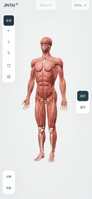
    • A fading title introduces two choices: whole body and organs on the left, bones, muscles and peripheral nerves on the right.
    • Review the selected model and usage notes, then press OK to begin.
  26. Published

    Left-side view and tool controls

    • Stacked view and rotation controls at the upper left. Explode and system buttons at the lower left open bottom panels.
  27. Published

    Reorganized the menu and explanation layout

    • Moved view controls into one scrollable Menu with a vertical layout.
    • Kept Select and Hide modes on the side, with explanations below a separate model viewport.
  28. Published

    Collapsible viewing controls

    • Small edge tabs reveal model, search and auxiliary controls. Interacting with the model hides them.
    • Moved the explosion slider into an optional disclosure while keeping selection, focus and undo accessible.
  29. Published

    Animated focus zoom

    • Smoothly zoom toward the selected part over 0.65 seconds while preserving the viewing direction.
    • Direct camera interaction interrupts the transition. Reduced-motion preferences skip the animation.
  30. Published

    Direct focus and undo controls

    • Added Focus to selection actions to isolate, enlarge and rotate selected parts, with an exit back to the whole model.
    • After hiding parts, show Undo with the remaining action count and Restore all directly on the scene.
  31. Published

    Fade out hidden parts

    • Hidden parts fade out gently over 0.32 seconds in both models.
    • Hidden parts stop receiving clicks immediately. Undo restores them immediately, and reduced-motion preferences skip the animation.
  32. Published

    Added a full, scrollable update history

    • Listed all recorded changes, newest first.
    • Count updates automatically, separately from the initial version and research records.
    • Keep the heading visible while scrolling through the full history.
  33. Published

    Corrected mirrored surface orientation

    • Fixed mismatches between triangle winding and lighting normals in mirrored Z-Anatomy parts.
    • Corrected 547 parts, including the left plantar aponeurosis, while preserving positions and triangle membership.
    • Validated all 1,138 parts and versioned the geometry to avoid stale cached data.
  34. Published

    Refined the selection colors

    • Changed warm-part highlights to peacock blue and cool-part highlights to rose.
    • Reduced the glow and slowed the pulse to approximately 2.8 seconds.
  35. Published

    Removed the floor for views from underneath

    • Removed the floor, platform and decorative rings.
    • Expanded the vertical orbit range to allow viewing the soles from below.
  36. Published

    Show three actions when selecting a part

    • Stopped opening the explanation automatically when a part is selected.
    • Show Details, Hide parts and Clear selection buttons.
    • Open the existing explanation panel only when Details is pressed, in both models.
  37. Published

    Made selected parts easier to identify

    • Added blue highlights for warm-colored parts and magenta for cool-colored parts.
    • Added a gentle 2.2-second pulse, disabled when reduced motion is requested.
    • Localized close-button labels, the glossary heading and the Z-Anatomy source description.
  38. Research

    Japanese translation audit

    • Reviewed 2,137 untranslated name records, representing 1,455 distinct English labels, with candidates and unresolved questions.
    • Classified 608 note fields containing Latin characters, including untranslated text, abbreviations, names and notation.
    • This records research, not a completed or published translation of all terms.
  39. Published

    Added Z-Anatomy and peripheral limb nerves

    • Added 1,138 parts, including skeletal, muscular and connective structures and 178 peripheral limb nerve parts.
    • Added model switching and a comparison of coverage, use cases and licenses.
    • Added Select/Hide modes, individual hiding, undo and restore for both models.
    • Added a desktop-only guide to rotation, panning and zoom.
    • Added an in-app update history.
  40. Date unknownPublished

    Initial atlas: explore the whole body

    • Included 2,234 BodyParts3D parts, 3,432 searchable concepts and 15 systems.
    • Added orbit, zoom, pan, preset views and automatic rotation.
    • Added system visibility, search, isolation and an exploded part inventory.
    • Added Japanese/English switching, a bilingual glossary, and general/clinical explanations. Some names remain untranslated.
    • Adapted panels for mobile and added source credits, licenses and usage information.